How Can I Correct Cell Types That Are Not Present In The Liver
April 23, 2022
Over the past few days, some readers have reported encountering cell types not found in the liver.
Recommended: Fortect
Introduction
The liver is usually one of the largest organs in the body and also plays a critical role in the metabolism of prescription drugs. Liver disease causes more than 2 million deaths worldwide each year, of which 1 million are complications of liver cirrhosis and 1 million are due to viral liver diseases and hepatocellular carcinoma (Asrani et’s. , 2019). Creating the right simulation model is critical in preclinical drug development and disease research. However, species-specific enzymes and drug-metabolizing transporters (DMETs) involved in drug uptake, distribution, metabolism, and excretion alter the drug’s pathway of action and prevent the use of animal tools for predicting toxicity in humans (Olsonotheral., Cheung 2000; and Gonzalez, 2008 ). Since then, it has been found that traditional uniformly exposed 2D monolayers help signal and nutrient transmission, rather than interaction between cells and matrix cells. Ta , an accelerated and dedifferentiated loss of the wifi phenotype was observed in a 2D model of basic human hepatocytes, manifested by a completely new low level of expression of basic DMETs and a decrease in albumin production (Roweotheral., 2013). The earliest disturbances at this level of transcripts in primary hepatocytes were initially observed after 30 minutes, more and more about 4000 transcripts were differentially expressed during the first 24 hours of culture, which strongly affected the pathways involved in the citric acid cycle, not to mention oxidative phosphorylation. from the synthesis of urea. (Lauschke et al., 2016).
Introduction
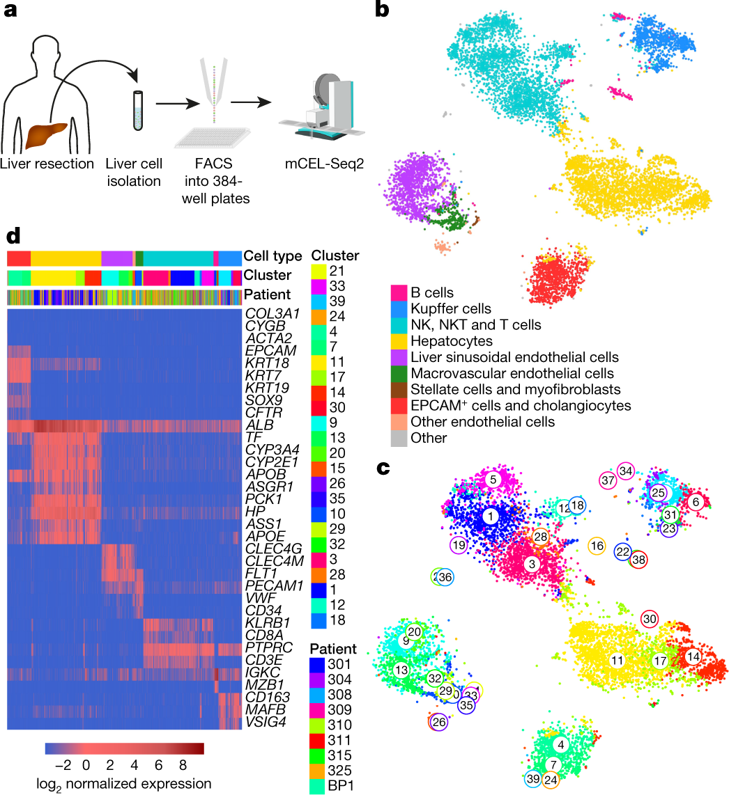
A hard-working liver is essential for human metabolic function. A reference map of the landscape of a healthy human liver, available at single cell resolution, is critical to unraveling the pathogenesis and treatment of diseases of the renal system. This landscape has been tedious to describe1, primarily because access to fresh liver tissue from a real person is limited and it is difficult to fractionate tissue to fractionate damage without Fragile resident cell communities. One approach to building a tolerant map of the human liver cell landscape is to combine very careful separation of relatively large segments of healthy healthy individuals with single-cell RNA (scRNA-seq) sequencing. While scRNA-seq is a good, robust, and powerful tool for describing the highly heterogeneous cell populations found in human tissues2,3, it is far from being useful for describing entire human organs, as only human cell roadmap islands extracted from i look like human pancreas published so far 4,5,6,7,8,9,10,11. Currently, the only single-cell Google transcriptome map covers the entire mouse liver12.
Liver Organoids
Liver Organoids are intelligent 3D in vitro models of my liver, as a platform for tales of the various wonders of research related to liver development and regeneration of your body, metabolic research, modeling of liver disease, and adult chemistry and biology.sound stem cells. Liver organelles, composed of round, unilamellar bay epithelium, support important physiological functions of the liver and are used to isolate and expand hepatic checkpoints and progenitor cells from stem cell niches.
>
The Liver Transcriptome
/h2> Transcriptome Analysis Behind The Liver Can Be Visualized By Looking At The Specificity And Distribution Of Transcribed MRNA Molecules (Figure 1). Specificity Measures How Many Of Your Genes Are Highly Or Poorly Expressed Only In The Liver Compared To Other Individuals. Overexpression Includes Three Sub-categories Of Overexpression:
Intrahepatic Cholangiocarcinoma (bile Duct Cancer)
About 10% or 20% of these cancers starting in each of our livers are intrahepatic cholangiocarcinomas. These cancers begin in the cells that line the small bile ducts (tubes that carry bile to the gallbladder) of the liver. However, most cholangiocarcinomas begin in the bile ducts of the liver.
HOME
LiverIt is a very important organ in the human body. It serves as a central metabolic manager with many very important functions, including the regulation of glucose and lipid metabolism, protein synthesis, bile activity and secretion, and the biotransformation of metabolites and xenobiotics. In addition, one type of liver is a visceral organ capable of significant natural renewal after tissue loss1. However, the prevalence and mortality of liver disease has increased dramatically in recent decades, with hepatocellular carcinoma (HCC) becoming the second leading cause of cancer death2,3. One of the main reasons for the rejection of effective treatments for a wide range of liver diseases is the complication of the organ and the abundance of little-studied cell types in it, which contribute to the pathogenesis of health problems. The cellular landscape of my liver has rarely been studied at single cell resolution, severely limiting our molecular understanding of liver function and disease biology. A serious problem was also the difficulty of isolating good viable genes.atocytes from patient tissues.
Identification Of Type-specific Transcriptional Portraits Of Defective Liver Cells Under Normal Biological Conditions
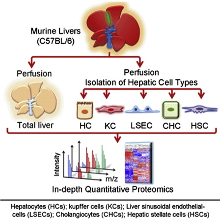
To characterize the client molecules that are HEPs. To present HSCs and/or ECs under physiological conditions, we used three transgenic lines Tg(fabp10a:dsRed), Tg(hand2:EGFP) such as Tg(kdrl:Hsa.HRAS-mCherry) expressing red (dsRed, mCherry) or green fluorescent proteins (GFP) by the respective cell types [14, 20, 23]. Whole livers were excised from adult zebrafish of each of their transgenic strains used in this training process (Figure 1A). Liver fluorescence microscopy of the respective transgenic lines confirmed their observed fluorescence in identical cell types (Fig. 1B). We prepared cell knockdown and performed FACS according to previously established protocols (see Methods, Appendix, Fig. 1). The number of scans of RNA sequences that actually matched fluorescent reporters that were almost cell type specific (Figure 1B) were heavily overpopulated in fluorescent positive samples, This, as you can see, confirmed the purity of the isolated FACS (Figure 1C). samples). To determine the exact genetic signatures of cell types, we performed differential predictive comparisons between samples and identified the most enriched genes in each cell type (Fig. 2A, Appendix Table 2). Most cell-specific genes were available in EC (4553), followed by HSC (380) and HEP (126) (Table 2 in the Appendix). These include well-known cell-specific paintball markers for EC (sox18 [24], sele [25], [26]), flt1, and HEP (soat2 [27]) (Figure 2B). On the other hand, transferred Doe genes are associated with fatty acids, metabolic processes (fasn [28], fat3b, hmgcra [29], hmgcs1 [30], elovl4a [31]) and cholesterol biosynthesis (cyp51, sc5d, hmgcra, msmo1, nsdhl , hmgcs1, dhcr7) are activated in HSCs known to contain vitamin A fat globules [32] (Supplementary Table 2). Gene ontology (GO) analysis revealed enrichment of angiogenesis-related genes in ECs, insulin-like growth factor receptor signaling your genes, and cellular phosphate ion homeostasis in HEP and lipid transport and exercise genes in HSCs (Figure 2C). . Together Enrichment of well-known markers, such as the corresponding GO terms in EC, HEP and HSC, maintains the individuality of the respective cell types.
Recommended: Fortect
Are you tired of your computer running slowly? Is it riddled with viruses and malware? Fear not, my friend, for Fortect is here to save the day! This powerful tool is designed to diagnose and repair all manner of Windows issues, while also boosting performance, optimizing memory, and keeping your PC running like new. So don't wait any longer - download Fortect today!

Hepatitis B Vaccination
may reduce your risk of hepatitis B Reduce your risk Hepatitis B vaccine. The vaccine can be given to almost anyone, including infants, the elderly, and immunosuppressed people.
Definitions
Cytology is the name given to the branch of biology that studies cells from their products, structure and functions.[1] Liver cytology specializes in liver cells. However, the main cells of the liver are called hepatocytes; There are other cells that can be seen in a liver sample, namely Kupffer cells (macrophages) [2]. Lean meat is the largest gland in the body. It performs a wide range of functions, ranging from the destruction of old blood cells to the control of the overall metabolism of macromolecules.[3] In the fetus, the liver usually acts as the main concentrate of hematopoiesis, the function of which is subsequently replaced by the bone marrow. This hematopoietic function is usually not observed afterLe birth; however, under certain pathological conditions, this feature may still be visible. It is important to note that the liver is a necessary organ and the only organ in the body that has the ability to regenerate immediately after surgery or injury.[4]
Celtypes Worden Niet Gevonden In De Lever
Tipy Kletok Ne Obnaruzheny V Pecheni
Les Types De Cellules Ne Se Trouvent Pas Dans Le Foie
I Tipi Cellulari Non Si Trovano Nel Fegato
간에서 세포 유형이 발견되지 않음
Tipos De Celulas No Se Encuentran En El Higado
Typy Komorek Nie Wystepuja W Watrobie
Celltyper Finns Inte I Levern
Tipos De Celulas Nao Sao Encontrados No Figado
Zelltypen Werden Nicht In Der Leber Gefunden